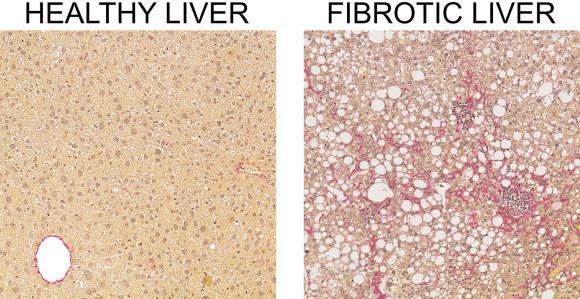
Closing in on liver fibrosis: Detailing the fibrosis process at unprecedented resolution
Today, there is no effective way to treat liver fibrosis. In a new study, researchers from University of Southern Denmark present a new technology to investigate the cellular processes as they change during fibrosis development. Key findings are being validated in studies of human patients, paving the way for possible novel diagnostics and treatments.
The human liver carries out a wealth of vital functions through highly coordinated processes involving multiple cell types. However, when the liver is damaged by pharmaceuticals, cholestasis or chronic fat accumulation caused by alcohol or metabolic dysfunction, the various cell types undergo pathological changes and liver function deteriorates. Extensive inflammation severely affects most cellular processes and scar tissue (liver fibrosis) gradually replaces normal liver tissue.
The global prevalence of both liver inflammation and liver fibrosis has taken on pandemic proportions, but still no effective treatment is available. Liver fibrosis may hence develop into cirrhosis and liver cancer with liver transplantation as last resort.

A much better understanding of the cellular dynamics in the healthy and fibrotic liver is therefore essential for the development of diagnostic tools and therapies to timely diagnose and reverse advanced liver fibrosis.
Watch the liver cell changes as they happen
In this project, researchers from University of Southern Denmark used mouse models of liver fibrosis and single-cell RNA sequencing technology to investigate the cellular processes as they change during fibrosis development.
At unprecedented resolution, they have detailed the fibrotic process and the changing interactions between liver cell types. Importantly, top genes found to correlate with liver fibrogenesis in mice turned out to be very accurate in the diagnosis also of human fibrosis.
Key findings are now being validated in studies of human patients aimed at further exploring novel diagnostic markers and identifying possible molecular targets for pharmacological intervention in the fibrotic process.
This validation is part of a larger study, conducted by the Center for Functional Genomics and Tissue Plasticity of patients undergoing bariatric surgery. For that study, the researchers are studying liver biopsies taken before and 18 months after surgery to understand, which cell types and genes change and how they change.
Authors and support
The findings have been published in the journal Hepatology. Main authors are graduate student Mike K. Terkelsen and Head of Research, Kim Ravnskjær from Department of Biochemistry and Molecular Biology, University of Southern Denmark. Contributing authors are from same department and Department of Molecular Medicine, Department of Clinical Research, Department of Pathology, Department of Gastroenterology and Hepatology, and Department of Nephrology, University of Southern Denmark.
This work was supported by the National Danish Research Foundation, the Fuhrmann Foundation, the Independent Research Fund Denmark, the European Commissions’ Marie Sklodowska-Curie Action, the European Foundation for the Study of Diabetes and University of Southern Denmark.
Meet the researcher
Kim Ravnskjær, Associate Professor, is head of a research group studying the development and resolution of liver fibrosis and non-alcoholic steatohepatitis in in the context of obesity and diabetes.