New technology could revolutionise treatment of osteoarthritis and cartilage injuries
Researchers from King's College London, the University of Southern Denmark, and Boston University have further developed a laser imaging technology that, for the first time ever, allows for holistic deep imaging of living tissue without damaging it. The study has just been published in Nature Communications.
Eight out of ten Danes develop osteoarthritis after the age of 50. The primary cause of this disease is damage to cartilage tissue, which cannot regenerate itself.
In severe cases, with the help of so-called tissue engineering, new cartilage can be cultivated in a laboratory and transplanted into the joint, especially the knee. However, since it is an expensive and cumbersome process, it is reserved for very few patients.
This may now change.
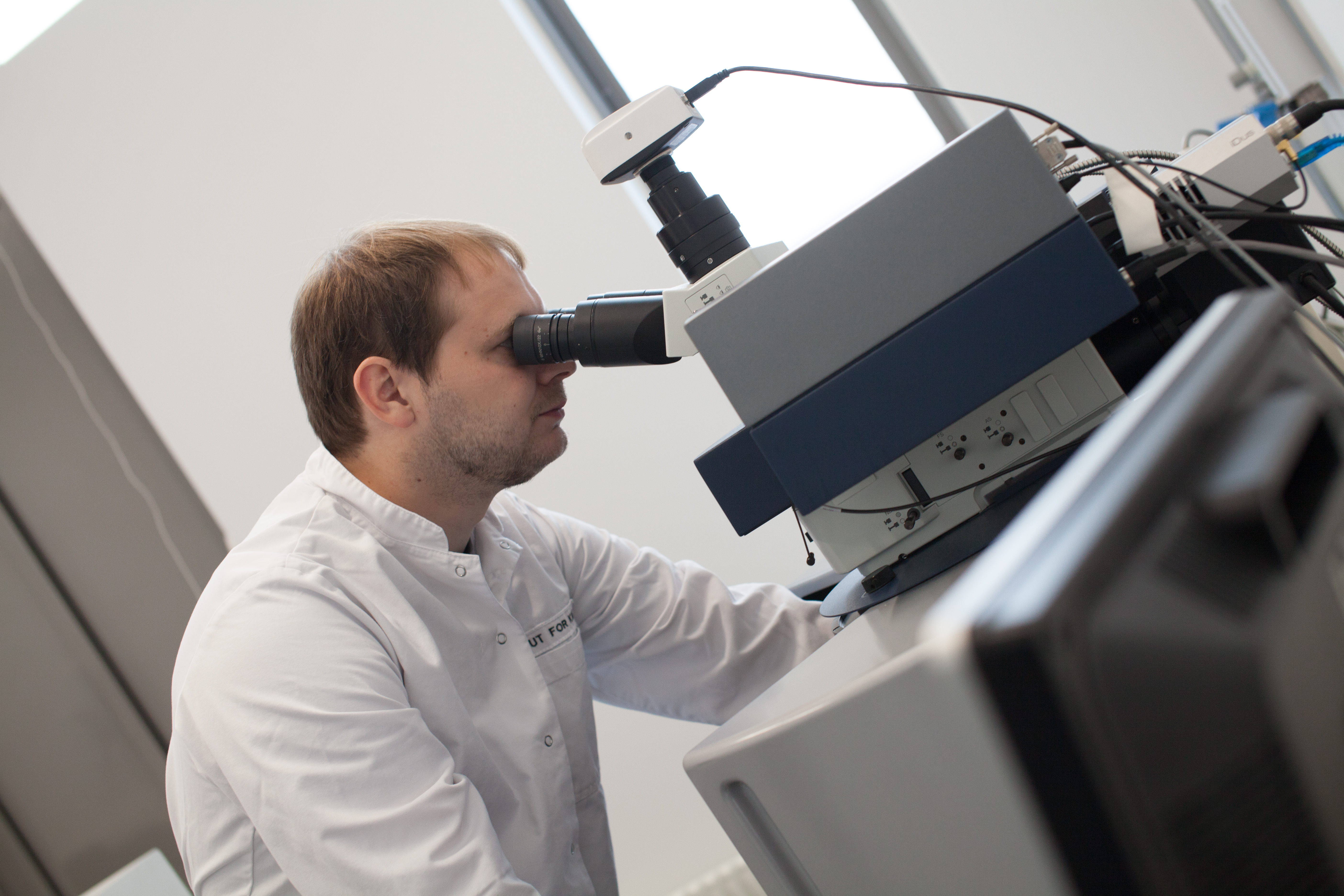
Researchers from King’s College London, the University of Southern Denmark, and Boston University have just published a study in Nature Communications on a further development of Raman spectroscopy technology, which uses laser light to analyse molecules in a substance. The researchers have succeeded in using the method to take pictures deep inside living tissue.
And this is a significant advance, they write, because it means that with Raman spectroscopy, one can now examine large pieces of tissue without damaging them.
According to the researchers, this discovery could therefore be used to promote the spread of tissue technology and halve the costs of cultivating cartilage in a laboratory.
As it stands now, one challenge with tissue technology is that two sets of cartilage must be produced. One set is examined to ensure it has been made correctly and is safe to transplant into the body, and another set for the actual transplantation. This is necessary because current methods for examining tissues use fluorescent substances that destroy the tissue.
Another problem is that one cannot be entirely sure if the two produced pieces of tissue are actually identical, even though they are produced in the same way.
All this will be solved by the new technology because one would only need one set of cartilage, which both undergoes examination and transplantation.
And the researchers behind the study see many more potential applications for the technology beyond just cartilage transplantation.
For example, they hope it can be used surgically in operating rooms when removing tumours. This would reduce the need for time-consuming pre-examinations of tumours, which could ultimately save lives.
- This system opens up numerous possibilities for imaging tissue, not only in relation to tissue technology but also in the treatment of cancer and autoimmune diseases, says Martin Hedegaard, Associate Professor of Biophotonics at the University of Southern Denmark and one of the authors of the study.
- For the first time, Raman is not primarily a surface-sensitive technology. Now we can see through a larger amount of tissue and obtain information about where specific signals originate. This will be valuable when investigating the spread of cancer and the development of autoimmune diseases such as rheumatoid arthritis.
The researchers are currently working on commercialising the technology and creating a spinout company with support from Spinouts Denmark.